|

|
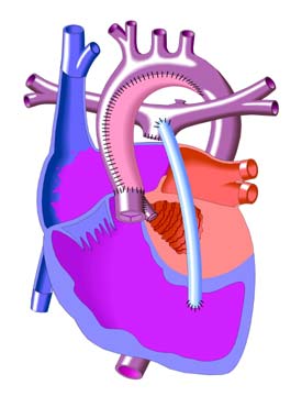
Sano Modification Procedure
The Sano Modification of the Norwood involves the placement of a conduit (light blue tube below) between the pulmonary artery and the right ventricle instead of the Modified Blalock-Taussig Shunt.
Sano Procedure
The first operation serves to make the right ventricle the main pumping chamber for blood flow to the body. The aorta is made larger to increase blood flow to the body. A connection is made to enable the blood traveling through the aorta towards the body to "shunt" through this connection and flow into the pulmonary artery to receive oxygen. This may be accomplished with a modified Blalock-Taussig shunt, which is a small tube placed between the aorta and the pulmonary arteries or by using the Sano modification procedure, in which a homograft (tissue) conduit is placed between the right ventricle and the pulmonary arteries. The choice of which procedure is best for your child can be discussed with your cardiologist and/or cardiovascular surgeon. However, even after the stage I procedure, the infant will still have some degree of cyanosis since oxygen-poor (blue) blood from the right atrium and oxygen-rich (red) blood from the left side of the heart mix and flow through the aorta to the body.
The Sano Shunt
Over the last few years, a number of centers around the world have begun to adopt a modification of the Norwood procedure that involves a different type of shunt. Introduced by Shunji Sano, MD, who was trained in congenital cardiac surgery in Melbourne, Australia, and is now working in his home country of Japan, this new modification indicates further improvement in the survival of newborn babies with hypoplastic left heart syndrome.
The Sano shunt modification avoids the problem of competitive flow between the lungs and coronary arteries. The shunt is constructed from a slightly larger Gortex tube graft than that used for the modified Blalock shunt. Generally a 5 mm tube graft is selected in contrast to the 3.5 mm graft used for averagesize babies for a Blalock shunt. Distally, the graft is connected to the main pulmonary artery between the right and left pulmonary artery takeoffs. The proximal end of the shunt is connected to a limited infundibular incision in the right ventricle.
|